EYE EXAM EQUIPMENT

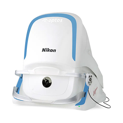
Optos Monaco
The Optos Monaco is an advanced retinal imaging system that combines ultra-widefield photography with OCT technology, allowing doctors to capture detailed images of up to 200° of the retina in a single scan. This enables earlier detection and more precise monitoring of eye diseases, all in a fast, non-invasive exam.
OptiLift
The OptiLift is a non-invasive treatment designed to improve dry eye by strengthening the eyelid muscles and enhancing blink function. Using gentle muscle stimulation and radiofrequency, it helps promote better tear distribution while also tightening and rejuvenating the skin around the eyes.
Firefly Slit Lamp
A slit lamp camera system is a medical device used to examine the eye. It combines a slit lamp biomicroscope, which provides a magnified view of the eye, with a digital camera that captures high-resolution images of the eye. The system allows us to observe the eye in detail, including the cornea, lens, and retina, and diagnose various eye conditions such as cataracts, glaucoma, macular degeneration, and diabetic retinopathy. We can then document and demonstrate these abnormalities of the eye to our patients.

Auto Lensmeter
Automated lensometry is designed to quickly and accurately determine the prescription of eyeglass lenses. Advanced technology allows us to analyze the lens and provide precise measurements of the lens power and other parameters. Overall, automated eyeglass lens reading devices are an important tool for optometrists and other eye care professionals. They help to streamline the process of determining the correct prescription for patients, and they help to ensure that patients receive the most accurate and effective eyeglass lenses possible.

Optical Response Analyzer
Our new optical response analyzer is a crucial tool in detecting potential for glaucoma.

OCULUS Keratograph 5M
This Keratograph allows us to provide dry eye evaluations at a higher level of accuracy than traditional methods.

TearLab Osmolarity System
The TearLab Osmolarity System is intended to measure the osmolarity of human tears to aid in the diagnosis of dry eye disease in patients suspected of having dry eye disease, in conjunction with other methods of clinical evaluation. TearLab is for professional in vitro diagnostic use only.
Hyperosmolarity has been described in the literature as a primary marker of tear film integrity. When the quantity or quality of secreted tears is compromised (known as aqueous deficient or evaporative Dry Eye Disease), increased rates of evaporation lead to a more concentrated tear film (increased osmolarity) that places stress on the corneal epithelium and conjunctiva.

iZon
Patients who are wearing new iZon Lenses report that their vision is clearer, sharper and more vivid than ever before. iZon Lenses have also been clinically proven to improve nighttime driving vision by dramatically reducing glare and providing better definition. In FDA-validated tests, iZon wearers were able to recognize and react to a hazard in the road 25 feet sooner than conventional lens wearers. Call our office to see if you are a candidate for iZon High Resolution Lenses. Click here to view more information on iZon’s website.

Optos Daytona
Our office features the top-of-the-line Optos Daytona to get an ultra-widefield view of the retina (the back of the eye). While eye exams generally include a look at the front of the eye to evaluate health and prescription changes, a thorough screening of the retina is critical to verify that your eye is healthy. This can lead to early detection of common diseases, such as macular degeneration, glaucoma, diabetes, and even cancer. The exam is quick, painless, and may not require dilation drops. To read about various conditions of the retina, click here.

Zeiss Cirrus HD-OCT
Some views draw the observer directly into the picture – views such as those offered by Cirrus HD-OCT. This new high-performance OCT instrument from Carl Zeiss Meditec offers a quantum leap forward. Featuring spectral domain technology, Cirrus HD-OCT delivers exquisite high-definition images of the ocular structures. Advanced optics aid in the examination of patients with cataracts. Dilation is not required even for pupils as small as 2.5 mm. Mouse Driven Alignment™ delivers superior image capture and analysis in just a few clicks, resulting in reduced chair time for the patient.

Pachymetry
Pachymeters play a vital role in glaucoma screening, the co-management of refractive surgery patients, and assessing corneal pathology. Pachymetry can be defined as the measurement of corneal thickness. Sound waves are bounced off tissues that form echoes, which are used to measure the corneal thickness.

Scout Wave Topographer
The Scout Topographer offers corneal topography, visible light pupillometry and dark-adapted pupillometry all on the same map. In addition, the Scout’s corneal wavefront maps clearly display lower-and higher-order aberrations that can be individually removed from the map for better understanding of problems. It is even possible to design a custom contact lens utilizing custom Wave Software. This is especially helpful in patients with post-surgical complications from RK and Corneal transplants.

Specular Microscopy
Specular microscopy enumerates the number and relative health of endothelial cells, allowing an objective assessment of the patient’s cornea. In cases of Fuchs’ corneal dystrophy, the cornea begins to show swelling and the vision becomes blurred. Specular microscopy is used to measure the corneal endothelium cell density and determine the presence of corneal swelling. For more information, click here.

Octopus 1-2-3 Visual Field Instrument
Determines peripheral and central vision disorders.

DGH 8000 ScanMate
The Scanmate is a self- contained, portable, multiple mode ultrasound imaging system. The Scanmate user can rapidly obtain high-resolution video loops and still images with clear, sharp details. Post-processing of the images includes playback, gain adjustment, contrast and image intensity variation, zoom without distortion, and distance measurements with dual digital calipers. All these features help provide a better diagnosis and improve the quality of patient care.

Ophthalmoscope
Examines the internal portion of the eye for a wide range of problems.

Compound Microscope
Used to examine patient’s eyelashes for Demodex folliculorum and Demodex Brevis which are mites found in the hair follicles, especially the eyelashes. Older people are much more likely to carry the mites; estimates range as high as a 96-98% infestation rate in aged people. The lower rate of children may be due to the fact that children produce much less sebum. Our office looks for these mites by carefully removing an eyelash or eyebrow hair and placing it under a microscope.
Wheelchair Access
Our office and equipment are wheelchair accessible.


Services
Learn More
Keep
In Touch
Monday
8:30 AM to 5:00 PM
Tuesday
8:30 AM to 6:00 PM
Wednesday
8:30 AM to 5:00 PM
Thursday
8:30 AM to 5:00 PM
Friday
8:30 AM to 5:00 PM
Powered by 